by John R. Mangiardi, M.D. and Howard Kane, Wm.
The meningioma is the neurosurgeon's "friend" and often his most enduring challenge. For both the physician and patient, this tumor carries a true tag of benign. It also carries the possibility of "cure" in approximately 80% of cases. Thus, the long-term outcome for a patient with this tumor is a direct function of the skill and assiduousness of the surgeon who removes it.
Elsewhere in the Brain Surgery Information Center's Primer on Brain Tumor Biology, it was mentioned that "benign" often does not really mean benign. Be assured that in this case, the tumor really is benign.
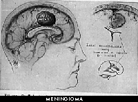
As mentioned earlier in the Primer, each type of brain tumor arises from a specific cell type. The cell of origin for the meningioma is call the arachnoid cap cell, found on the surface coverings (called meninges) of the brain in the paccionian granulations. These serve as the one-way valve system between the water system of the brain and the veins that drain from the brain to the heart.
Interestingly, these tumors have an embryologic relationship with cells found in the muscle layer of the utereus. In fact, it is exceedingly difficult for the pathologist to distinguish the meningioma from the fibroid tumors of the utereus under the microscope. Also, they share the characteristic female hormonal receptors (estrogen and progesterone) on their cell surfaces. This characteristic has lead to the testing of anti-estrogen receptor agents, such as tamoxifin, as a growth-inhibiting agent in these tumors. Clinical studies to date have failed to provide siginificantly positive results.
Meningiomas are rarely malignant in their behavior. But when malignant, meningiomas grow rapidly and are destructive; they are quite difficult to treat, and recur oftentimes in less than a year after surgical removal. They are also difficult for the pathologist to diagnose under the microscope. Probably the only finding that correlates well with the diagnosis is that of numerous cells seen in division ("mitosis"). The pathologist may occasionally speak of brain and skull invasion, cells with an abnormal appearance, or other bizarre findings, however none of these completey fit the diagnosis. Ultimately, the diagnosis is determined by the activity of the particular tumor over time.
A cousin to the meningioma is the hemangiopericytoma. The cell of origin for this tumor is the perivascular pericyte (located around blood vessels). Although very similar to the benign meninigiomas, these tumors tend to recur with great rapidity (less than one year) and frequency. Some physicians classify these tumors with the malignant meningiomas.
0 Comments:
Post a Comment